

In The Above Animal Cell, What Is The Function Of The Cellular Organelle Labeled With The Letter Y?
| Organelle | |
|---|---|
| Details | |
| Pronunciation | |
| Role of | Prison cell |
| Identifiers | |
| Latin | Organella |
| MeSH | D015388 |
| Thursday | H1.00.01.0.00009 |
| FMA | 63832 |
| Anatomical terms of microanatomy [edit on Wikidata] | |
In cell biology, an organelle is a specialized subunit, usually within a cell, that has a specific function. The proper name organelle comes from the idea that these structures are parts of cells, as organs are to the body, hence organelle, the suffix -elle beingness a diminutive. Organelles are either separately enclosed inside their ain lipid bilayers (likewise called membrane-bound organelles) or are spatially distinct functional units without a surrounding lipid bilayer (non-membrane spring organelles). Although most organelles are functional units within cells, some functional units that extend outside of cells are often termed organelles, such as cilia, the flagellum and archaellum, and the trichocyst.
Organelles are identified past microscopy, and can also be purified past cell fractionation. At that place are many types of organelles, peculiarly in eukaryotic cells. They include structures that brand upwards the endomembrane system (such as the nuclear envelope, endoplasmic reticulum, and Golgi apparatus), and other structures such as mitochondria and plastids. While prokaryotes do not possess eukaryotic organelles, some do comprise protein-shelled bacterial microcompartments, which are thought to act as primitive prokaryotic organelles;[1] and there is also testify of other membrane-bounded structures.[2] Also, the prokaryotic flagellum which protrudes outside the cell, and its motor, besides as the largely extracellular pilus, are oft spoken of as organelles.
History and terminology [edit]
| Prison cell biological science | |
|---|---|
| Animal cell diagram | |
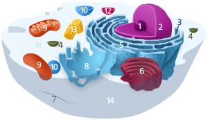 Components of a typical creature cell:
|
In biology organs are defined as confined functional units within an organism.[3] The analogy of bodily organs to microscopic cellular substructures is obvious, as from fifty-fifty early on works, authors of respective textbooks rarely elaborate on the distinction betwixt the two.
In the 1830s, Félix Dujardin refuted Ehrenberg theory which said that microorganisms have the same organs of multicellular animals, but pocket-size.[4]
Credited as the first[five] [6] [7] to use a diminutive of organ (i.e., picayune organ) for cellular structures was German zoologist Karl August Möbius (1884), who used the term organula (plural of organulum, the diminutive of Latin organum).[viii] In a footnote, which was published as a correction in the next outcome of the journal, he justified his suggestion to call organs of unicellular organisms "organella" since they are only differently formed parts of one jail cell, in contrast to multicellular organs of multicellular organisms.[viii] [9]
Types [edit]
While nigh cell biologists consider the term organelle to be synonymous with cell compartment, a space ofttimes leap past ane or ii lipid bilayers, some cell biologists cull to limit the term to include but those cell compartments that contain deoxyribonucleic acid (DNA), having originated from formerly autonomous microscopic organisms acquired via endosymbiosis.[x] [11] [12]
Nether this definition, at that place would only be two broad classes of organelles (i.e. those that comprise their own Dna, and have originated from endosymbiotic bacteria):
- mitochondria (in virtually all eukaryotes)
- plastids[13] (eastward.one thousand. in plants, algae, and some protists).
Other organelles are likewise suggested to take endosymbiotic origins, simply do not contain their ain DNA (notably the flagellum – see evolution of flagella).
A second, less restrictive definition of organelles is that they are membrane-bound structures. However, even by using this definition, some parts of the cell that take been shown to be distinct functional units practice non authorize as organelles. Therefore, the use of organelle to as well refer to non-membrane jump structures such equally ribosomes is mutual and accustomed.[fourteen] [15] [16] This has led many texts to delineate between membrane-bound and non-membrane bound organelles.[17] The non-membrane jump organelles, besides called big biomolecular complexes, are large assemblies of macromolecules that carry out item and specialized functions, just they lack membrane boundaries. Many of these are referred to as "proteinaceous organelles" every bit their main structure is made of proteins. Such cell structures include:
- large RNA and protein complexes: ribosome, spliceosome, vault
- large poly peptide complexes: proteasome, DNA polymerase III holoenzyme, RNA polymerase II holoenzyme, symmetric viral capsids, complex of GroEL and GroES; membrane protein complexes: porosome, photosystem I, ATP synthase
- large Dna and protein complexes: nucleosome
- centriole and microtubule-organizing eye (MTOC)
- cytoskeleton
- flagellum
- nucleolus
- stress granule
- germ cell granule
- neuronal transport granule
The mechanisms by which such non-membrane bound organelles form and retain their spatial integrity take been likened to liquid-liquid stage separation.[18]
Eukaryotic organelles [edit]
Eukaryotic cells are structurally complex, and by definition are organized, in office, by interior compartments that are themselves enclosed by lipid membranes that resemble the outermost jail cell membrane. The larger organelles, such as the nucleus and vacuoles, are easily visible with the light microscope. They were among the beginning biological discoveries made after the invention of the microscope.
Not all eukaryotic cells have each of the organelles listed below. Exceptional organisms have cells that do not include some organelles that might otherwise be considered universal to eukaryotes (such every bit mitochondria).[19] At that place are likewise occasional exceptions to the number of membranes surrounding organelles, listed in the tables below (e.g., some that are listed equally double-membrane are sometimes found with single or triple membranes). In addition, the number of individual organelles of each blazon institute in a given cell varies depending upon the function of that cell.
| Organelle | Primary function | Structure | Organisms | Notes |
|---|---|---|---|---|
| jail cell membrane | separates the interior of all cells from the outside environment (the extracellular space) which protects the cell from its environment. | 2-dimensional liquid | all eukaryotes | |
| cell wall | The jail cell wall is a rigid structure composed of cellulose that provides shape to the cell, helps continue the organelles within the jail cell, and does not let the cell flare-up from osmotic pressure level. | diverse | plants, protists, rare kleptoplastic organisms | |
| chloroplast (plastid) | photosynthesis, traps energy from sunlight | double-membrane compartment | plants, protists, rare kleptoplastic organisms | has ain DNA; theorized to be engulfed by the ancestral eukaryotic jail cell (endosymbiosis) |
| endoplasmic reticulum | translation and folding of new proteins (rough endoplasmic reticulum), expression of lipids (smoothen endoplasmic reticulum) | single-membrane compartment | all eukaryotes | rough endoplasmic reticulum is covered with ribosomes, has folds that are flat sacs; smooth endoplasmic reticulum has folds that are tubular |
| flagellum | locomotion, sensory | protein | some eukaryotes | |
| Golgi apparatus | sorting, packaging, processing and modification of proteins | single-membrane compartment | all eukaryotes | cis-face (convex) nearest to crude endoplasmic reticulum; trans-face (concave) farthest from rough endoplasmic reticulum |
| mitochondrion | energy production from the oxidation of glucose substances and the release of adenosine triphosphate | double-membrane compartment | most eukaryotes | constituting element of the chondriome; has own DNA; theorized to take been engulfed by an ancestral eukaryotic cell (endosymbiosis)[20] |
| nucleus | Dna maintenance, controls all activities of the jail cell, RNA transcription | double-membrane compartment | all eukaryotes | contains bulk of genome |
| vacuole | storage, transportation, helps maintain homeostasis | single-membrane compartment | eukaryotes |
Mitochondria and plastids, including chloroplasts, have double membranes and their own Dna. Co-ordinate to the endosymbiotic theory, they are believed to take originated from incompletely consumed or invading prokaryotic organisms.
| Organelle/Macromolecule | Primary office | Structure | Organisms |
|---|---|---|---|
| acrosome | helps spermatozoa fuse with ovum | single-membrane compartment | nigh animals |
| autophagosome | vesicle that sequesters cytoplasmic textile and organelles for deposition | double-membrane compartment | all eukaryotes |
| centriole | anchor for cytoskeleton, organizes cell division by forming spindle fibers | Microtubule poly peptide | animals |
| cilium | move in or of external medium; "disquisitional developmental signaling pathway".[21] | Microtubule poly peptide | animals, protists, few plants |
| cnidocyst | stinging | coiled hollow tubule | cnidarians |
| eyespot apparatus | detects calorie-free, allowing phototaxis to take place | green algae and other unicellular photosynthetic organisms such equally euglenids | |
| glycosome | carries out glycolysis | unmarried-membrane compartment | Some protozoa, such as Trypanosomes. |
| glyoxysome | conversion of fatty into sugars | single-membrane compartment | plants |
| hydrogenosome | energy & hydrogen production | double-membrane compartment | a few unicellular eukaryotes |
| lysosome | breakdown of big molecules (e.g., proteins + polysaccharides) | single-membrane compartment | animals |
| melanosome | pigment storage | single-membrane compartment | animals |
| mitosome | probably plays a role in Iron–sulfur cluster (Iron–S) assembly | double-membrane compartment | a few unicellular eukaryotes that lack mitochondria |
| myofibril | myocyte contraction | bundled filaments | animals |
| nucleolus | pre-ribosome production | protein–DNA–RNA | most eukaryotes |
| ocelloid | detects calorie-free and possibly shapes, allowing phototaxis to take identify | double-membrane compartment | members of the family Warnowiaceae |
| parenthesome | not characterized | not characterized | fungi |
| peroxisome | breakdown of metabolic hydrogen peroxide | unmarried-membrane compartment | all eukaryotes |
| porosome | secretory portal | single-membrane compartment | all eukaryotes |
| proteasome | degradation of unneeded or damaged proteins past proteolysis | very large protein complex | all eukaryotes, all archaea, and some leaner |
| ribosome (80S) | translation of RNA into proteins | RNA-protein | all eukaryotes |
| stress granule | mRNA storage[22] | membraneless (mRNP complexes) | virtually eukaryotes |
| TIGER domain | mRNA encoding proteins | membraneless | virtually organisms |
| vesicle | material transport | single-membrane compartment | all eukaryotes |
Other related structures:
- cytosol
- endomembrane organisation
- nucleosome
- microtubule
Prokaryotic organelles [edit]
Prokaryotes are not as structurally complex every bit eukaryotes, and were once thought as having fiddling internal organization, and lack cellular compartments and internal membranes; but slowly, details are emerging about prokaryotic internal structures that overturn these assumptions.[2] An early on false turn was the idea developed in the 1970s that leaner might contain cell membrane folds termed mesosomes, but these were subsequently shown to be artifacts produced by the chemicals used to prepare the cells for electron microscopy.[24]
However, there is increasing evidence of compartmentalization in at least some prokaryotes.[2] Recent research has revealed that at least some prokaryotes accept microcompartments, such as carboxysomes. These subcellular compartments are 100–200 nm in diameter and are enclosed by a beat of proteins.[1] Even more than hit is the description of membrane-spring magnetosomes in bacteria, reported in 2006.[25] [26]
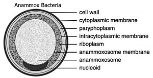
The bacterial phylum Planctomycetota has revealed a number of compartmentalization features. The Planctomycetota prison cell plan includes intracytoplasmic membranes that separates the cytoplasm into paryphoplasm (an outer ribosome-costless space) and pirellulosome (or riboplasm, an inner ribosome-containing space).[27] Membrane-jump anammoxosomes have been discovered in v Planctomycetota "anammox" genera, which perform anaerobic ammonium oxidation.[28] In the Planctomycetota species Gemmata obscuriglobus, a nucleus-like structure surrounded by lipid membranes has been reported.[27] [29]
Compartmentalization is a feature of prokaryotic photosynthetic structures.[two] Purple bacteria accept "chromatophores", which are reaction centers institute in invaginations of the prison cell membrane.[2] Green sulfur bacteria have chlorosomes, which are photosynthetic antenna complexes found bonded to cell membranes.[2] Cyanobacteria have internal thylakoid membranes for calorie-free-dependent photosynthesis; studies take revealed that the prison cell membrane and the thylakoid membranes are non continuous with each other.[ii]
| Organelle/macromolecule | Master function | Construction | Organisms |
|---|---|---|---|
| anammoxosome | anaerobic ammonium oxidation | ladderane lipid membrane | "Candidatus" bacteria within Planctomycetota |
| carboxysome | carbon fixation | poly peptide-shell bacterial microcompartment | some bacteria |
| chlorosome | photosynthesis | light harvesting complex fastened to cell membrane | green sulfur leaner |
| flagellum | move in external medium | protein filament | some prokaryotes |
| magnetosome | magnetic orientation | inorganic crystal, lipid membrane | magnetotactic bacteria |
| nucleoid | Dna maintenance, transcription to RNA | Dna-protein | prokaryotes |
| pilus | Adhesion to other cells for conjugation or to a solid substrate to create motile forces. | a hair-like bagginess sticking out (though partially embedded into) the plasma membrane | prokaryotic cells |
| plasmid | Deoxyribonucleic acid exchange | circular Dna | some leaner |
| ribosome (70S) | translation of RNA into proteins | RNA-protein | bacteria and archaea |
| thylakoid membranes | photosynthesis | photosystem proteins and pigments | mostly blue-green alga |
Encounter also [edit]
- CoRR hypothesis
- Ejectosome
- Endosymbiotic theory
- Organelle biogenesis
- Membrane vesicle trafficking
- Host-pathogen interface
- Vesiculo-vacuolar organelle
References [edit]
- ^ a b Kerfeld CA, Sawaya MR, Tanaka S, Nguyen CV, Phillips M, Beeby M, Yeates TO (Baronial 2005). "Protein structures forming the beat out of archaic organelles". Science. 309 (5736): 936–8. Bibcode:2005Sci...309..936K. CiteSeerX10.1.1.1026.896. doi:x.1126/science.1113397. PMID 16081736. S2CID 24561197.
- ^ a b c d e f g Murat, Dorothee; Byrne, Meghan; Komeili, Arash (2010-ten-01). "Jail cell Biology of Prokaryotic Organelles". Cold Leap Harbor Perspectives in Biological science. 2 (ten): a000422. doi:10.1101/cshperspect.a000422. PMC2944366. PMID 20739411.
- ^ Peterson L (Apr 17, 2010). "Mastering the Parts of a Cell". Lesson Planet. Retrieved 2010-04-xix .
- ^ Di Gregorio MA (2005). From Here to Eternity: Ernst Haeckel and Scientific Faith. Gottingen: Vandenhoeck & Ruprecht. p. 218.
- ^ Bütschli O (1888). Dr. H. Thou. Bronn'south Klassen u. Ordnungen des Thier-Reichs wissenschaftlich dargestellt in Wort und Bild. Erster Band. Protozoa. Dritte Abtheilung: Infusoria und System der Radiolaria. p. 1412.
Die Vacuolen sind demnach in strengem Sinne keine beständigen Organe oder O r g a n u l a (wie Möbius die Organe der Einzelligen im Gegensatz zu denen der Vielzelligen zu nennen vorschlug).
- ^ Ryder JA, ed. (February 1889). "Embryology: The Structure of the Human Spermatozoon". American Naturalist. 23: 184.
It may perchance be of advantage to employ the give-and-take organula here instead of organ, following a suggestion by Möbius. Functionally differentiated multicellular aggregates in multicellular forms or metazoa are in this sense organs, while, for functionally differentiated portions of unicellular organisms or for such differentiated portions of the unicellular germ-elements of metazoa, the diminutive organula is advisable.
- ^ Robin C, Pouchet G, Duval MM, Retterrer East, Tourneux F (1891). Journal de fifty'anatomie et de la physiologie normales et pathologiques de fifty'homme et des animaux. F. Alcan.
- ^ a b Möbius K (September 1884). "Das Sterben der einzelligen und der vielzelligen Tiere. Vergleichend betrachtet". Biologisches Centralblatt. 4 (13, 14): 389–392, 448.
Während die Fortpflanzungszellen der vielzelligen Tiere unthätig fortleben bis sie sich loslösen, wandern und entwickeln, treten die einzelligen Tiere auch durch die an der Fortpflanzung beteiligten Leibesmasse in Verkehr mit der Außenwelt und viele bilden sich dafür auch besondere Organula". Footnote on p. 448: "Die Organe der Heteroplastiden bestehen aus vereinigten Zellen. Da die Organe der Monoplastiden nur verschieden ausgebildete Teile e i n e r Zelle sind schlage ich vor, sie „Organula" zu nennen
- ^ Walker, Patrick (2009). Nuclear import of histone fold motif containing heterodimers by importin thirteen. Niedersächsische Staats-und Universitätsbibliothek Göttingen.
- ^ Keeling PJ, Archibald JM (Apr 2008). "Organelle evolution: what's in a name?". Current Biology. 18 (8): R345-7. doi:10.1016/j.cub.2008.02.065. PMID 18430636. S2CID 11520942.
- ^ Imanian B, Carpenter KJ, Keeling PJ (March–April 2007). "Mitochondrial genome of a tertiary endosymbiont retains genes for electron transport proteins". The Journal of Eukaryotic Microbiology. 54 (ii): 146–53. doi:10.1111/j.1550-7408.2007.00245.x. PMID 17403155. S2CID 20393495.
- ^ Mullins C (2004). "Theory of Organelle Biogenesis: A Historical Perspective". The Biogenesis of Cellular Organelles. Springer Science+Business Media, National Institutes of Health. ISBN978-0-306-47990-8.
- ^ Alberts B, Johnson A, Lewis J, Raff M, Roberts One thousand, Walter P (2002). "The Genetic Systems of Mitochondria and Plastids". Molecular Biology of the Prison cell (4th ed.). ISBN978-0-8153-3218-3.
- ^ Campbell NA, Reece JB, Mitchell LG (2002). Biology (6th ed.). Benjamin Cummings. ISBN978-0-8053-6624-2.
- ^ Nott TJ, Petsalaki E, Farber P, Jervis D, Fussner E, Plochowietz A, Craggs TD, Bazett-Jones DP, Pawson T, Forman-Kay JD, Baldwin AJ (March 2015). "Phase transition of a disordered nuage protein generates environmentally responsive membraneless organelles". Molecular Prison cell. 57 (5): 936–947. doi:ten.1016/j.molcel.2015.01.013. PMC4352761. PMID 25747659.
- ^ Banani SF, Lee HO, Hyman AA, Rosen MK (May 2017). "Biomolecular condensates: organizers of cellular biochemistry". Nature Reviews Molecular Jail cell Biology. 18 (5): 285–298. doi:10.1038/nrm.2017.7. PMC7434221. PMID 28225081.
- ^ Cormack DH (1984). Introduction to Histology . Lippincott. ISBN978-0-397-52114-2.
- ^ Brangwynne CP, Eckmann CR, Courson DS, Rybarska A, Hoege C, Gharakhani J, Jülicher F, Hyman AA (June 2009). "Germline P granules are liquid droplets that localize by controlled dissolution/condensation". Science. 324 (5935): 1729–32. Bibcode:2009Sci...324.1729B. doi:ten.1126/science.1172046. PMID 19460965. S2CID 42229928.
- ^ Fahey RC, Newton GL, Arrick B, Overdank-Bogart T, Aley SB (April 1984). "Entamoeba histolytica: a eukaryote without glutathione metabolism". Science. 224 (4644): lxx–2. Bibcode:1984Sci...224...70F. doi:x.1126/science.6322306. PMID 6322306.
- ^ Alberts B, Johnson A, Lewis J, Morgan D, Raff MC, Roberts Yard, Walter P, Wilson JH, Hunt T (2014-11-eighteen). Molecular biology of the cell (Sixth ed.). Garland Science. p. 679. ISBN978-0815345244.
- ^ Badano JL, Mitsuma North, Beales PL, Katsanis N (September 2006). "The ciliopathies: an emerging grade of human genetic disorders". Annual Review of Genomics and Man Genetics. 7: 125–48. doi:ten.1146/annurev.genom.seven.080505.115610. PMID 16722803.
- ^ Anderson P, Kedersha N (March 2008). "Stress granules: the Tao of RNA triage". Trends in Biochemical Sciences. 33 (3): 141–50. doi:10.1016/j.tibs.2007.12.003. PMID 18291657.
- ^ Tsai Y, Sawaya MR, Cannon GC, Cai F, Williams EB, Heinhorst S, Kerfeld CA, Yeates TO (June 2007). "Structural assay of CsoS1A and the protein crush of the Halothiobacillus neapolitanus carboxysome". PLOS Biological science. 5 (6): e144. doi:x.1371/periodical.pbio.0050144. PMC1872035. PMID 17518518.
- ^ Ryter A (January–February 1988). "Contribution of new cryomethods to a better knowledge of bacterial anatomy". Annales de l'Institut Pasteur. Microbiology. 139 (1): 33–44. doi:10.1016/0769-2609(88)90095-6. PMID 3289587.
- ^ Komeili A, Li Z, Newman DK, Jensen GJ (January 2006). "Magnetosomes are jail cell membrane invaginations organized by the actin-like poly peptide MamK" (PDF). Science. 311 (5758): 242–5. Bibcode:2006Sci...311..242K. doi:x.1126/science.1123231. PMID 16373532. S2CID 36909813.
- ^ Scheffel A, Gruska M, Faivre D, Linaroudis A, Plitzko JM, Schüler D (March 2006). "An acidic poly peptide aligns magnetosomes along a filamentous structure in magnetotactic leaner". Nature. 440 (7080): 110–4. Bibcode:2006Natur.440..110S. doi:10.1038/nature04382. PMID 16299495. S2CID 4372846.
- ^ a b Lindsay, Grand. R.; Webb, R. I.; Strous, M; Jetten, M. S.; Butler, M. K.; Forde, R. J.; Fuerst, J. A. (2001). "Jail cell compartmentalisation in planctomycetes: Novel types of structural organisation for the bacterial cell". Archives of Microbiology. 175 (6): 413–29. doi:10.1007/s002030100280. PMID 11491082. S2CID 21970703.
- ^ Jetten, Mike S. K.; Niftrik, Laura van; Strous, Marc; Kartal, Boran; Keltjens, Jan T.; Op den Camp, Huub J. Thou. (2009-06-01). "Biochemistry and molecular biological science of anammox bacteria". Disquisitional Reviews in Biochemistry and Molecular Biological science. 44 (ii–3): 65–84. doi:ten.1080/10409230902722783. PMID 19247843. S2CID 205694872. Retrieved 2020-08-03 .
- ^ Fuerst JA (Oct 13, 2005). "Intracellular compartmentation in planctomycetes". Annual Review of Microbiology. 59: 299–328. doi:10.1146/annurev.micro.59.030804.121258. PMID 15910279.
External links [edit]
-
 Media related to Organelles at Wikimedia Commons
 - Tree of Life project: Eukaryotes
- Organelle Databases
Source: https://en.wikipedia.org/wiki/Organelle
Posted by: reimereaketury.blogspot.com
0 Response to "In The Above Animal Cell, What Is The Function Of The Cellular Organelle Labeled With The Letter Y?"
Post a Comment